Bacteria Have a Secret Engineering Trick to Keep Themselves in Shape
NYU scientists discover that the bacterial cell wall exploits an unusual mechanical non-linearity to grow at a constant width — a discovery that may also apply to plant roots
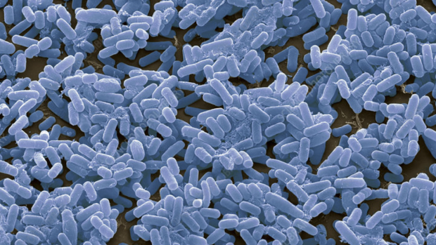
Bacillus subtilis, a rod bacteria.
Blow up a long balloon and two things happen: it gets longer and it gets wider.
Now imagine a living cell that inflates itself under enormous pressure and yet only grows longer, never adding width. That is exactly what rod-shaped bacteria do, every time they divide, with a precision that has baffled scientists for decades.
A new study published in Current Biology has finally found the answer. Researchers suggest their discovery could point toward new treatments for antibiotic-resistant bacteria.
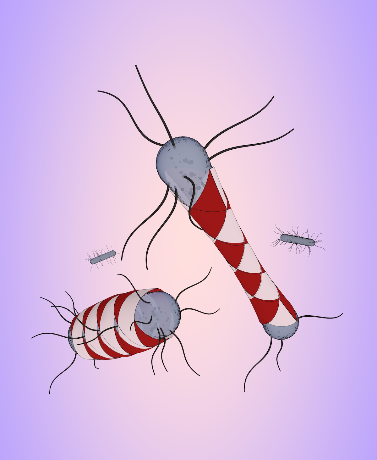
Rod-shaped bacteria like Bacillus subtilis — a harmless soil microbe and one of biology's most studied model organisms — are encased in a rigid shell called the cell wall, made of a polymer called peptidoglycan, and pressurized from within at many times the pressure of a car tire.
To grow, bacteria must continuously remodel this wall: snipping out old material with enzymes and weaving in new polymer. This should cause the cell to bulge outward as well as elongate. Yet rod-shaped bacteria hold their width to within 40 nanometers — roughly 1,750 times thinner than a human hair.
"Most antibiotics that target the bacterial cell wall disrupt its structure/architecture," said Paola Bardetti, the study's lead author and an industry assistant professor of chemical and biomolecular engineering at NYU Tandon School of Engineering. "Our work reveals an entirely different vulnerability: the physical mechanism bacteria rely on to maintain their shape. No drug has ever targeted that. Until now, we didn't understand it well enough to try."
The NYU team, led by Bardetti and the paper’s senior author Enrique Rojas, an associate professor of biology at NYU, subjected living bacteria to rapid osmotic shocks — briefly raising or lowering internal pressure — while tracking wall deformations as small as a few nanometers.
What they found was a sharp mechanical threshold. Below normal pressure the wall behaves like a finger trap toy: reducing pressure makes it expand sideways. Above it, the wall softens and the cell widens. At the transition, width stays constant, precisely where growing bacteria sit.
“The cell wall is a smart material,” said Bardetti. “It responds to mechanical stress in a way that is tuned to keep the cell the right shape. Every time we probed it, it surprised us.”
This tipping-point strategy also confers automatic self-correction. When cells were manipulated to grow wider than normal, the wall slipped into the finger-trap regime, thinning it back toward the target width. The critical pressure of the transition also shifted in response to changes in wall architecture — a second feedback loop — making this a homeostatic system encoded in the physical properties of the material itself.
In scientific terms: the wall is anisotropic, far stiffer circumferentially than longitudinally, with a Poisson ratio of 0.45–0.5 and anisotropy at the physical maximum. The stress-softening non-linearity — an abrupt drop in circumferential stiffness at the critical pressure — parks the cell at the boundary between widening and thinning.
The same phenomenon appeared in Arabidopsis thaliana plant roots, suggesting a shape-control strategy evolution has arrived at independently.
"Finding the same strategy in bacteria and plant roots was genuinely exciting," said Bardetti. "It suggests a fundamental principle of tubular morphogenesis that nature has independently discovered more than once. The next step is identifying the molecular machinery that sets the critical pressure, because once you know that, you have a potential drug target.”
In addition to Bardetti and Rojas, the paper’s authors are Felix Barber (currently Assistant Professor at Ohio State University) and Dylan Fitzmaurice, postdoctoral researcher and PhD candidate respectively in the Rojas Lab at the time of the study. Research funding came from the National Institutes of Health and the National Science Foundation. Microscopy support was provided by the NYU Langone Health Microscopy Lab, partially funded by the National Cancer Institute.
Non-linear stress-softening of peptidoglycan mediates bacterial cell shape homeostasis
Bardetti, Paola et al. Current Biology, Volume 36, Issue 5, 1156 - 1165.e5