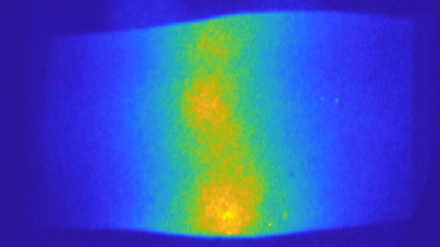
Evaluation of lupus arthritis using frequency domain optical imaging

Example of a frequency-domain image of a finger joint (proximal interphalangeal joint of index finger) affected by lupus arthritis.
Systemic lupus erythematosus (SLE), commonly referred to as simply “lupus”, is an autoimmune disorder where the body’s immune system attacks healthy tissue. Lupus affects somewhere between 20 to 150 people per 100,000, with variations among different racial and ethnic groups. The disease often causes arthritic symptoms in the joints, which can be debilitating in some cases.
Despite the severity of the disease, identification of lupus arthritis and assessment of its activity remains a challenge in clinical practice. Evaluations based on traditional joint examination lack precision, due to its subjective nature and accuracy in situations such as obese digits and co-existing fibromyalgia. As such, these examinations have limited ability to render quantitative data about improvement and worsening.
Recently, imaging technology, especially ultrasound (US) and magnetic resonance imaging (MRI), has enabled more objective and detailed assessment of articular and periarticular abnormalities with higher sensitivity. However, MRI and US are expensive and time-consuming. Furthermore, US has been found to be very operator dependent. Therefore, both modalities are currently not routinely used in practice. There is a clear unmet need for a simple, reliable, non-invasive and low-cost imaging modality that can objectively assess and monitor arthritis progress in patients with lupus.
Now, researchers at NYU Tandon in collaboration with Columbia University are exploring optical imaging technology as a reliable way to diagnose patients and assess the progression of the disease. The researchers, including Research Assistant Professor Alessandro Marone and Chair of the Biomedical Engineering Department Andreas H. Hielscher, found that frequency domain optical imaging could reliably identify lupus arthritis, and could be used to track how the disease progressed.
The results provide strong evidence that frequency domain optical images could provide objective, accurate insights into SLE that were not possible or economically feasible using other technologies. The light diffusion identified inflammation in the blood vessels around joints, similar to but distinct from the symptoms caused by rheumatoid arthritis. With this technology, caregivers may not have to rely on patient feedback to track the progression of lupus, but can see it in action.
Optical imaging methods have been used in studies comparing osteoarthritis, rheumatoid arthritis (RA) and healthy controls. The results of those studies highlighted that patients suffering from RA have higher light absorption in the joint space compared with healthy subjects. This is likely due to the presence of inflammatory synovial fluid that decreases light transmission through the inflamed joints. But these observations have never been used to study lupus before, and these findings could provide a reliable, rapid, and cost-effective method of assessing joint involvement in lupus patients.


