
NYU Engineers and Surgeons Funded to Build a Genetic Test Fast Enough for the Operating Room
A $3.3 million NIH grant will help NYU Tandon and NYU Langone researchers automate a rapid test that could identify brain tumor type and remaining cancer cells within minutes
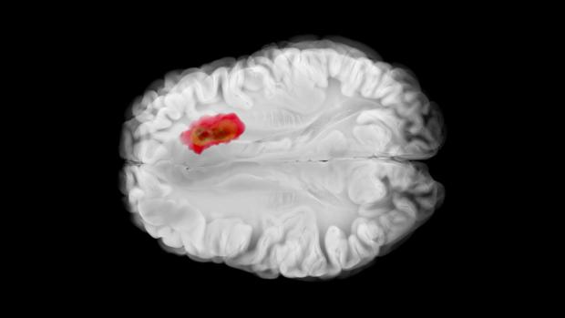
Engineers and brain surgeons at NYU are developing a genetic testing device that could tell surgeons, within minutes, what type of glioma brain tumor they are removing and whether cancer cells remain at its edges.
Gliomas are a relatively common type of brain tumor that infiltrates surrounding brain tissue. As a result, surgeons must balance removing as much tumor as possible against the risk of damaging healthy parts of the brain.
The mutations a tumor carries also help determine its diagnosis, prognosis and treatment, but results typically arrive days or weeks after an operation.
"This technology will tell us about important aspects of a tumor’s genetics before the patient has even woken up from surgery," said Daniel Orringer, a neurosurgeon and co-director of the NYU Brain and Spine Tumor Center at NYU Langone Health's Perlmutter Cancer Center. "It could spare some patients a long, anxious wait and give surgeons information that shapes how they operate."
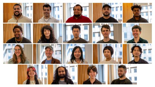
The project has received a five-year, $3.3 million grant from the National Cancer Institute, part of the National Institutes of Health. In addition to Orringer, the project is led by Katsuo Kurabayashi, chair of Mechanical and Aerospace Engineering (MAE) at NYU Tandon School of Engineering; Gilad Evrony, the Jacob D. Goldfield associate professor in NYU Langone Health's Center for Human Genetics & Genomics; and Yujing Song, Tandon MAE research assistant professor, as a co-investigator.
How completely a glioma is removed during surgery is an important predictor of a patient's outcome. The device would first look for mutations that are common in different types of brain tumors and then test tissue from the walls of the surgical cavity for mutations that are present in the tumor.
The test uses droplet digital PCR, or ddPCR, which divides a sample into thousands of microscopic droplets and copies targeted stretches of DNA inside them. Droplets containing a tumor mutation light up, allowing researchers to measure how much tumor DNA is present.
Even if run immediately, standard ddPCR takes about two to three hours, too long to be useful during an operation. Evrony's laboratory, working with Orringer, developed an ultra-rapid version that produces a result in about 15 minutes. In an initial study of 22 lower-grade glioma surgeries, it closely matched conventional ddPCR and could detect tumor cells making up as little as 0.1 percent of a sample.
"Our original version involves many manual steps and requires a very skilled technician," Evrony said. "In its current form, it’s not something that we could bring to other neurosurgery operating rooms."
Turning that manual process into an automated device is the job of Kurabayashi, a longtime developer of biomedical devices, several of them commercialized. Kurabayashi likens the division of labor to cooking. Evrony's lab developed the recipe, and Kurabayashi's team is building the machine that carries it out precisely and automatically.
In collaboration with Song, Kurabayashi is designing a device that can perform every step of the test for many samples at once in under 15 minutes.
"As engineers, getting our research out of the lab and into practical use is the whole point," Kurabayashi said. The researchers are partnering with Wainamics, a Silicon Valley company, which is helping design the device for inexpensive mass production. Bio-Rad is also providing reagents and instruments to support the project.
The initial focus is on lower-grade gliomas, which grow more slowly than glioblastoma, the most aggressive form, but can return and become more aggressive. Many carry mutations in genes called IDH1 and IDH2 that help classify the tumor.
Evrony will collaborate on developing the device and develop tests for a panel of mutations common in gliomas, so a single run at the start of surgery could identify which one a patient's tumor carries.
Orringer will lead a study of adults undergoing surgery at NYU Langone for suspected lower-grade gliomas, testing tissue from the tumor and its margins and comparing the results with MRI scans taken within 24 hours of surgery, the current standard for assessing whether tumor remains. During the study, results won't be shared with surgeons until after the operation, while the device's reliability is established.
"When people look and see an MRI looks clean," Evrony said, "is there actually tumor left behind that nobody knew about?"
By the end of the grant, the team aims to have a device ready for development in a form suitable for use in many brain tumor centers, a step the researchers hope will change how brain tumor surgery is done. The technology could eventually extend to other cancers and infectious diseases.
This project is supported by the National Cancer Institute of the National Institutes of Health under award number R01CA307127 and is funded 100 percent ($3.3 million) by federal funds, with no non-governmental funding. The content is solely the responsibility of the authors and does not necessarily represent the official views of the National Institutes of Health.