
Seeing More with Visible Light OCT: Structural and Spectral Imaging in Aging and AMD
Speaker:
Ruoyu Meng, PhD-Candidate
Dept. of Biomedical Engineering
NYU Tandon School of Engineering
Abstract:
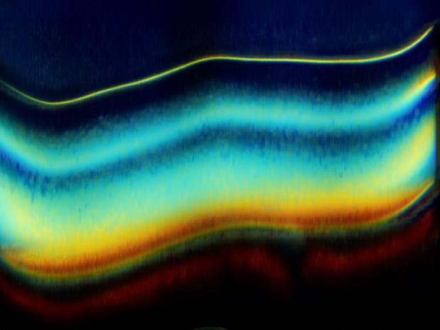
Age-related macular degeneration (AMD) is the leading cause of blindness in developed countries. With a rapidly aging global population, the prevalence and burden of AMD are expected to rise substantially. Because current treatments primarily target advanced stages, biomarkers that enable early diagnosis or predict disease progression are critically needed. Visible light OCT offers unique advantages over conventional near-infrared OCT, providing ultrahigh axial resolution and access to spectroscopic information within the visible spectrum. The improved resolution enables more precise delineation of retinal bands, each corresponding to specific cellular or subcellular structures. This finer detail facilitates detection of subtle changes associated with normal aging and early AMD, such as sub-micrometer alterations in the retinal pigment epithelium (RPE) and Bruch’s membrane (BM). Additionally, retinal chromophores exhibit characteristic extinction properties in the visible range; thus, chromophores such as macular pigment and RPE melanosomes can be quantified and visualized in vivo, offering enhanced biochemical contrast. Photoreceptors, responsible for phototransduction, can also be assessed more sensitively with visible-light OCT. Early irregularities-often obscured in near-infrared imaging-become detectable. While adaptive optics (AO) OCT enables high transverse-resolution optoretinography at the single-cell level, visible light OCT extends AO by providing depth-resolved structural information and spectroscopic changes in rod outer segments during stimulation, allowing direct evaluation of both structure and function. Advances and applications of visible light OCT are explored, including aging-related structural imaging, optoretinography, and densitometry. Expanding these capabilities to larger studies of AMD and other ocular diseases may accelerate the translation of visible light OCT into ophthalmic research and clinical care.
Ruoyu Meng earned a B.Eng. in Electronic Science and Engineering from Xidian University in 2017, and an M.Eng. in Biomedical Engineering from Tsinghua University, in 2020, where he worked on cancer detection using polarization imaging. He began his doctoral work in 2021 under the supervision of Prof. Vivek J. Srinivasan. He built a ultrahigh-resolution visible light OCT system in the Neurophotonics Lab at the Tech4Health Institute, NYU Langone Health. He conducted visible light OCT densitometry studies, visualizing and quantifying melanin within the retinal pigment epithelium (RPE) and macular pigment primarily in the Henle fiber layer (HFL) using spectroscopic contrast. He developed imaging protocols and analysis methods for visible light OCT-based optoretinography. Ruoyu has gained rich experience imaging patients referred from the clinic and initiated an ultrahigh-resolution human retina cohort. Based on this cohort, ongoing studies are focused on aging-related retinal structural changes and AMD characterization. In Summer 2025, Ruoyu completed an optical engineering internship at Triple Ring Technologies in California, working on optical sub-assemblies, laser beam characterization, and imaging system development.