A new way to track breast cancer
Researchers in the Clinical Biophotonics Laboratory are helping develop the technology to help doctors understand the development of cancerous tissue.

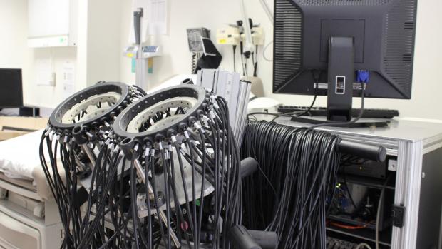
This optical tomography device that can be used to recognize and track breast cancer, without the negative effects of previous imaging technology. It uses near-infrared light to shine into breast tissue and measure light attenuation that is caused by the propagation through the affected tissue.
In the United States, roughly 13% of women will develop breast cancer, and more than 43,000 women are expected to die as a result in 2021. The disease represents a serious problem for public health. And while screening for the disease can help prevent catastrophic results, monitoring the progression of cancer and how it responds to treatment can be difficult.
Now researchers at NYU Tandon are developing the technology to help track the development of breast cancer, without causing further harm. And Mirella Altoe, Adjunct Professor of Biomedical Engineering and Postdoctoral Associate in the Clinical Biophotonics Laboratory, is helping lead this research.
Altoe grew up in Brazil, and attended the University of Brasilia as an undergraduate student in electrical engineering. While attending school, she found an opportunity to study abroad. She studied at UC Davis and John Hopkins, and did an internship at MIT. It was at MIT that she discovered her passion for biophotonics, the use of light to interact with body tissue in order to gleam information about biological processes and internal diseases. Altoe continued her program at MIT, then went back to Brazil to continue her undergraduate education.

“Studying in America, the opportunity to engage with this breakthrough research and learn from the professors that actually are developing these new technologies was the biggest reason behind my choice to study here” said Altoe.
The taste of biophotonics brought her back to the United States quickly. While at MIT, Altoe researched ways to improve the resolution field of surgical endoscopes, in order to better visualize the workings of the GI tract. Thanks to a scholarship from Brazil won from a national competition for young scholars, she applied for a Ph.D. program to study under Andreas Hielscher, currently the department chair at NYU Tandon and then at Columbia University. Hielscher’s lab was using a unique technology — optical tomography — to study breast cancer.
Altoe had previously done work at a hospital in Brazil, working on oncology across a wide range of cancers. The opportunity to continue that work — and to help develop a less invasive cancer monitoring system — appealed to her.
“Standard imaging modalities such as mammograms, ultrasound, or magnetic resonance imaging are mostly used to measure tumor size and are insensitive to tumor response,” said Altoe. “When it comes to treatment monitoring, these imaging modalities lack the versatility or efficacy that is needed to image patients multiple times safely at a reasonable cost.”
As a Ph.D. student, Altoe developed data processing and optical image analysis protocols to identify chemotherapy-induced changes in breast tissue metabolism in breast cancer patients. To accomplish this, she conducted a clinical study to collect data from more than 100 breast cancer patients undergoing chemotherapy. The research was designed to monitor changes in optically derived metrics from cancerous and healthy tissue. After earning her Ph.D., she officially joined the Clinical Biophotonics Laboratory under Hielscher.
Researchers in the lab are utilizing an optical tomography device that can be used to recognize and track breast cancer, without the negative effects of previous imaging technology. Traditional x-ray technology has a host of negative side effects due to radiation output, which makes it problematic to do regular scans on tumors. Their device uses near-infrared light to shine into breast tissue and measure light attenuation that is caused by the propagation through the affected tissue.
The change in the amplitude of the light as it passes through the breast is a result from water , lipids, and oxyhemogoblin’s distribution throughout the tissue, and they can use these as potential biomarkers as signs for cancerous tissue. By using the sensors, they can create 3D models of problematic tissue formations, and would be able to track tumors as they grow or shrink as treatments are applied. The technology, spearheaded by Hielscher, offers an opportunity for clinicians treating breast cancer patients to acquire far more information about their patients and their treatments than possible with other technologies.
The technology is not just limited to breast cancer. It has the potential to help study cancerous tissue in select other organs where light can be measured from end to end. It’s also being used to help diagnose other diseases, such as lupus.
Altoe began her work on this technology at Columbia, but followed Hielscher to NYU Tandon when he became the department chair of the school of Biomedical Engineering in 2020. Since then, she has published several papers in reputable journals related to the optical tomography technology and breast cancer treatment.
Altoe’s work has garnered her a host of recognitions, including an honorable mention for the ScientistA Award Recognizing Latina Women in Science, celebrating early career Brazilian women working both for academia and industry in the US. In addition to her research, she is teaching courses on the foundations necessary for understanding biological data analysis and how statistics can help extract scientific insights from data, laying the groundwork for a new generation of biomedical engineers.


