Diffusion MRI Nerve-Tract Visualization with Radial DSI and ODF-Fingerprinting
Speaker:
Steven Baete, PhD
Assistant Professor, Department of Radiology
NYU Grossman School of Medicine
Abstract:
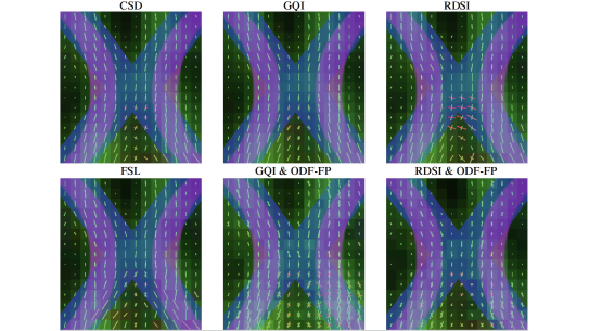
Dr. Baete and his team focuses on advancing magnetic resonance imaging (MRI) techniques for biomedical applications. Baete’s primary areas of expertise include dynamic diffusion imaging, diffusion spectrum imaging, and the development of novel MRI pulse sequences and acquisition methods. Through his work, he aims to improve the detection, characterization, and monitoring of various pathologies, with a particular emphasis on neurological disorders and cancer. One of Baete's key contributions is his research on orientation distribution function (ODF) fingerprinting, which enables improved reconstruction of crossing fibers in the mouse optic pathways using diffusion MRI. This technique has the potential to enhance our understanding of brain microstructure and connectivity, with applications in the study of neurological diseases. In addition to his methodological innovations, Baete collaborates extensively with clinicians to translate advanced MRI technologies into the clinic. He is actively involved in multidisciplinary research teams that leverage expertise from fields such as physics, engineering, and computer science to develop intelligent, multimodal imaging approaches. His contributions have the potential to significantly impact the field of biomedical imaging and improve patient care.
Dr. Baete received his Ph.D. in Life Sciences and Medicine from Ghent University, Belgium, in 2011. Subsequently he joined the Department of Radiology at NYU Langone Health as a Postdoctoral Fellow. He was appointed Assistant Professor of Radiology in 2017, and in 2021 became the Graduate Advisor for the Biomedical Imaging & Technology PhD Training Program at the Sackler Institute of Graduate Biomedical Sciences. Baete's work has been published in prestigious journals, and he is a highly respected member of the international MRI research community, serving on various committees and study groups.