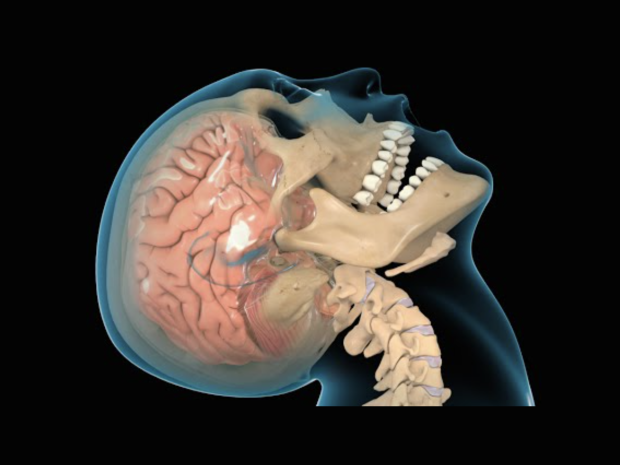
The Biomechanics of Traumatic Brain Injuries: Towards Better Prevention and Treatment
Speaker:
Bryan Pfister, Ph.D.
Professor and Chair
Department of Biomedical Engineering
New Jersey Institute for Technology
Abstract:
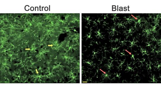
Dr. Pfister’s research encompasses how mechanical forces affect the nervous system – spanning from stretch induced growth during development to axonal stretch injury in traumatic brain injuries. He is also working to determine biomechanics of brain injury from blunt and blast injuries using both animate models and full-scale inanimate models of the human brain. He is also a co-PI on a grant from the Army to develop a primary-blast injury criteria for animal/human TBI models using field validated shock tubes. He is funded by the Army Research Laboratory, National Science Foundation, National Institutes of Health and the New Jersey Commission on Brain Injury Research.
Dr. Bryan Pfister received his BS from Clarkson University, earned his PhD in Material Science and Engineering from the Johns Hopkins University in 2002. Subsequently, he accepted a post-doctoral fellowship in the Department of Neurosurgery, University of Pennsylvania, and joined the NJIT Biomedical Engineering Department in January 2006, where he now serves as Department Chair. He played a leading role in the department’s initial ABET academic accreditation in 2006, and chaired the accreditation visit in 2013. More recently he has helped establish the Center for Injury Biomechanics, Materials and Medicine, New Jersey’s first Research Center focused on Nervous system injury and repair.