Quantitative ultrasound for musculoskeletal tissue imaging characteristics and therapeutic regeneration
Speaker:
Yi-Xian Qin, Ph.D.
Distinguished Professor and Chair
Department of Biomedical Engineering
Stony Brook University
Abstract:
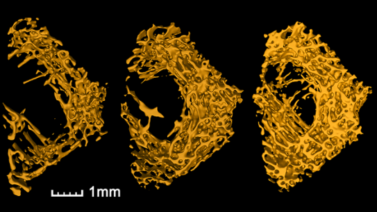
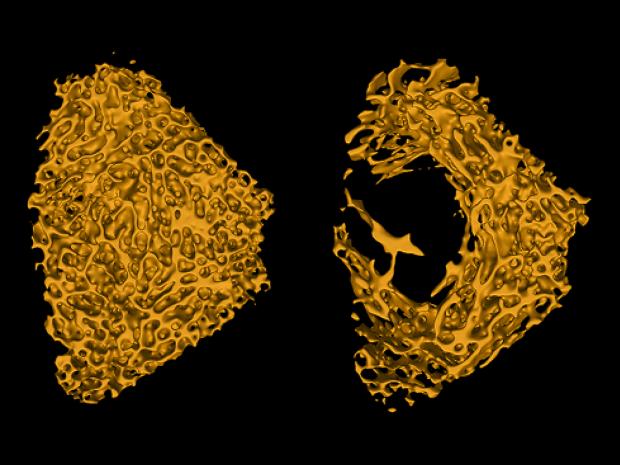
Dr. Qin is interested in the physical mechanisms involved in the control of tissue growth, healing, and homeostasis. In particular, he studies how bone adaptation and regeneration is influenced by the mechanical environment and how these mechanisms can be utilized in the treatment and prevention of disease and injury (e.g. osteoporosis and fracture healing). Bone senses and responds to biomechanical stimuli towards the achievement and maintenance of a structurally appropriate skeletal structure. Dr. Qin’s team investigates the interdependent roles of these mechanical signals through empiric and analytic models to provide support for the complex interactive mechanism of bone remodeling. In addition, they are developing acoustic diagnostic systems that allow them to image these effects and to improve tissue quality using the therapeutic aspects of ultrasound.
Dr. Qin received his PhD in Mechanical Engineering from SUNY Stony Brook in 1997. Subsequently he became a founding member of the Department of Biomedical Engineering at the same institution. Rising through the ranks he was named SUNY Distinguished Professor and Chair, Department of Biomedical Engineering in 2020. From 2018 through 2020, he held the position of Associate Dean for Academic Affairs and International Programs at the College of Engineering and Applied Sciences. Acknowledging the significance of his scientific contributions Dr. Qin has been elected fellow by several societies, including the American Society for Bone and Mineral Research (ASBMR), the American Society for Biomedical Engineering (BMES), the American Institute for Medical and Biological Engineering (AIMBE), and the International Academy of Astronautics (IAA)