Ultrafast MRI Diffusion Tensor Imaging for High Resolution Brain Mapping
Speaker:
Jiangyang Zhang, Ph.D.
Associate Professor, Department of Radiology
NYU Grossman School of Medicine
Abstract:
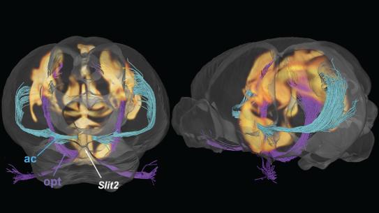
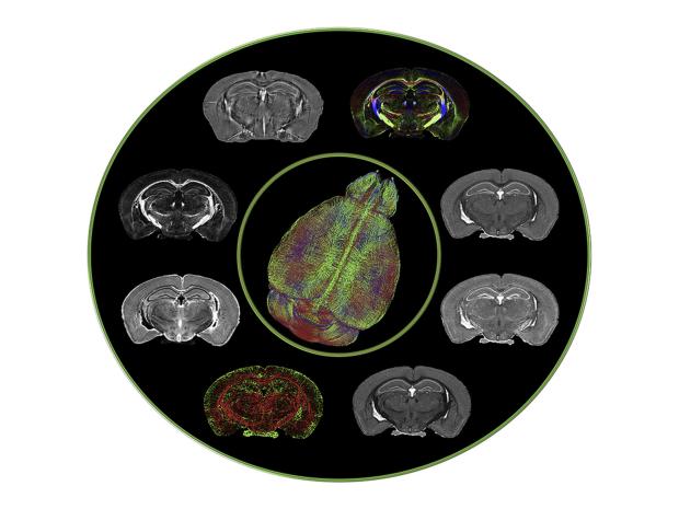
1H magnetic resonance imaging (MRI) maps brain structure and function non- invasively through versatile contrasts that exploit inhomogeneity in tissue micro- environments. Inferring histopathological information from MRI findings, however, remains challenging due to absence of direct links between MRI signals and cellular structures. In this talk we show that deep convolutional neural networks, developed using co- registered multi- contrast MRI and histological data of the mouse brain, can estimate histological staining intensity directly from MRI signals at each voxel. The results provide three- dimensional maps of axons and myelin with tissue contrasts that closely mimic target histology and enhanced sensitivity and specificity compared to conventional MRI markers. Further-more, the relative contribution of each MRI contrast within the networks can be used to optimize multi- contrast MRI acquisition. We anticipate our method to be a starting point for translation of MRI results into easy- to- understand virtual histology for neurobiologists and provide resources for validating novel MRI techniques.
Dr. Jiangyang Zhang started his academic career with a bachelor’s degree in electronic engineering from Tsinghua University in Beijing, China. The next 15 years he spent at Johns Hopkins University in Baltimore, Maryland. There he received an MSE degree in applied mathematics, and an MSE and PhD degree in Biomedical Engineering, before joining the faculty in the School of Medicine as an Assistant Professor in 2006. In 2013 he was promoted to Associate Professor, and in 2015 he accepted an offer from NYU’s Department of Radiology. He is an expert in diffusion MRI techniques that examine tissue microstructures as well as computational techniques to quantify the observed changes during development and injury. These techniques are useful to characterize abnormal brain development due to genetic mutations and structural abnormalities due to injuries.