
New imaging insights into osteoarthritis disease
Speaker:
Jose Maria M. Raya Garcia del Olmo, PhD
Associate Professor, Department of Radiology
NYU Grossman School of Medicine
Abstract:
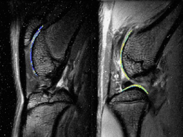
The complex and poorly understood etiology of the onset of posttraumatic osteoarthritis (PTOA) continues to hinder the development of effective therapeutic interventions to prevent progressive joint destruction after injury. Our ability to diagnose loss of tissue homeostasis will determine both how well we can phenotype PTOA and how effectively we can target therapeutic interventions. Molecular imaging has potential to target with high specificity a pathway overtime and identify tissues in which the pathway is upregulated. In molecular imaging, we exploit the specificity of a molecule (antibody, peptide, small molecule) to create an imaging agent by tagging it with a molecule that can be seen with at least one imaging modality (e.g., MRI). In this way, we can see where the molecules accumulate in the tissue in vivo. While molecular imaging has been extensively used in tumor, rheumatoid arthritis, and arteriosclerotic plaques, its application to OA has been anecdotal and its potential remains largely unexplored. In this talk,Dr. Raya will show how molecular imaging can be used to target and characterize pro-inflammatory events for knee PTOA. In particular, his teams assessed a promising peptide, P15, a 15-mer peptide that functions by binding hyaluronan (HA) and modulating its role in inflammatory response.
Dr. Raya received his PhD from the Ludwig Maximilian University (LMU) in Munich, Germany, in 2009. His thesis led to several publications, including topics such as diffusion-weighted imaging for bone tumors and acute vertebral fractures, validation of diffusion tensor imaging of articular cartilage, and methods for voxel-wise analysis of changes in articular cartilage. Subsequently he became a research scientist in NYU’s Department of Biomedical Engineering, where he focused on the translation of diffusion-weighted imaging to the musculoskeletal system. Receiving substantial external funding from National Institutes of Health (NIH) and the Arthritis Foundation, he was promoted to assistant professor in 2013 and associate professor in 2019
