Skeletal Tissue Regeneration and Engineering
Speaker:
Mitchell B. Schaffler, PhD
Professor and Chair, Dept. of Biomedical Engineering
The City College of New York (CCNY)
Abstract:
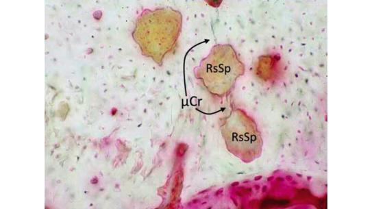
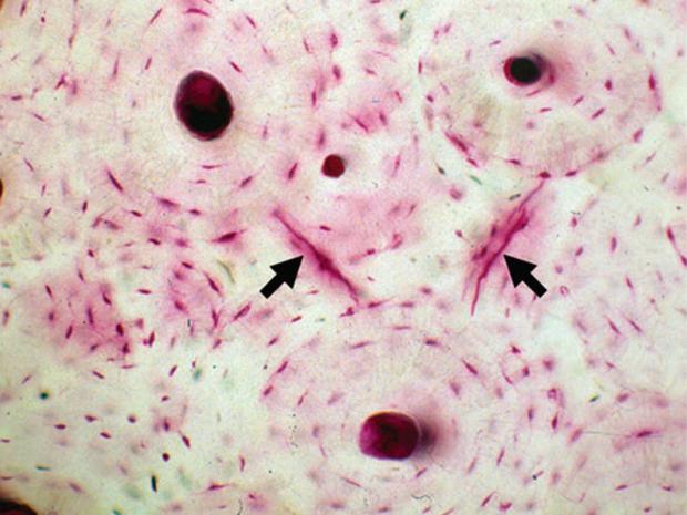
The major research emphasis of Dr. Schaffler’s Bone and Joint Laboratory (BJL) is to understand how skeletal tissues (bone, ligament, tendon, cartilage) develop, maintain and repair themselves in order to meet mechanical demands throughout life. Of particular interest are the cellular and integrative processes that control the architectural features of bone and tendon and how they govern the responses to physical challenges in aging and in diseases, such as osteoporosis, genetic defects and diabetes. Ongoing research efforts focus on fatigue and repair in bone and tendon, with specific emphasis on discovering how living cells in these tissues detect and repair wear and tear damage before it accumulates to the point of mechanical failure. Dr. Schaffler is also examining how osteocytes (the tissue-resident bone cells) influence s mechanical function, both directly by modulating local matrix composition, and indirectly by controlling local bone remodeling activities. Experimental approaches used in the BJL include in vivo and tissue mechanical loading studies, mechanical and biomaterials testing, microscopy, microcomputed tomography , in vivo microscopy and super-resolution microscopy, cell culture and molecular biology.
Dr. Schaffler received PhD degree in Anatomy & Orthopaedic Engineering from West Virginia. After being the Director of Orthopaedic Research at Mount Sinai School of Medicine, he joined CCNY in 2008. There he was named CUNY Distinguished Professor and Chair of the Department of Biomedical Engineering in 2012. Dr. Schaffler is an Elected Fellow of the American Institute for Medical and Biological Engineering, Elected Fellow, the American Society for Bone and Mineral Research, and the American Association of Anatomists. In addition, he received the AcroMed Award for Outstanding Research from the North American Spine Society. He has been a member of various journal editorial boards, including the Journal of Orthopaedic Research, BONE, and Anatomical Record; and served as reviewer for the
National Institute of Arthritis, Musculoskeletal and Skin Diseases (NIH), National Science Foundation, The Wellcome Trust (UK), and the Orthopaedic Research Society, among others.