Stem cell-based tissue engineering strategies to enhance bone regeneration

Speaker:
Philipp Leucht, MD
Associate Professor, Depts. of Orthopaedic Surgery and Cell Biology
NYU Grossman School of Medicine
Abstract:
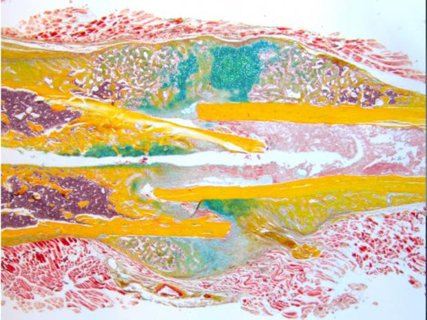
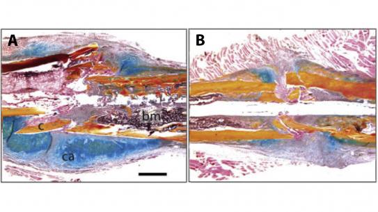
Many adult tissues harbor stem cells, which theoretically could be employed for injury repair, but the origins of these cells, and the factors that influence their developmental potency, are poorly understood. In his laboratory Dr. Leucht and his team address these questions in the context of skeletal tissue regeneration. He combines basic science and translational research and focuses on (1) determining the underlying cell and molecular regulatory mechanisms involved in skeletal development and fracture repair, (2) investigating the role of the mechanical environment during fracture repair, and (3) stem cell-based tissue engineering strategies to enhance bone regeneration. His group has developed in-vivo fracture healing models in mouse, rat and rabbit and employs bone histomorphometry, in situ hybridization, and microCT analysis. More recently he has explored a relatively new target indication: enhancing the osteogenic potential of aged bone autograft.
Dr. Leucht received his medical degree in 2002 at the Ruhr-University Bochum, Germany, which was followed by an orthopaedic residency at the Johann-Wolfgang-Goethe University in Frankfurt am Main. he started his clinician-scientist career in 2004, when he joined the laboratory of Dr. Jill Helms at Stanford University. There he finished his orthopedic residency and trauma fellowship and subsequently joined New York University Langone Medical Center. As an orthopaedic trauma-tologist, Dr. Leucht's long-term goal is to use the knowledge gained from basic research to improve fracture management.


