At the Crossroads: Engineering strategies for in vivo preclinical imaging at NYU

Speaker:
Youssef Wadghiri, PhD
Associate Professor of Radiology
Director of the Preclinical Imaging Laboratory
NYU Langone Health & NYU Grossman School of Medicine
Abstract:
To date all identified human disease associated genes can find their homologues in mice. It is these homologies that have made mouse models of disease the prevalent mammalian model of choice to study gene function and human disease. The research activities of the Wadghiri Lab are devoted to the development of non-invasive MRI strategies to facilitate the collection of anatomical and functional data of mouse models of human disease. The lab is also applying existing clinical protocols to the mouse in the areas of cancer and neurodegenerative diseases where MRI can be optimized and validated through a more precise correlation with histological data. In addition, his team is developing high-field MRI methods in combination with cellular and molecular targeting techniques to better understand human diseases and validate small animal imaging protocols that will be transferable to humans.
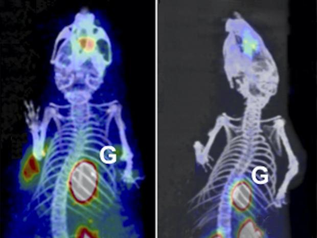
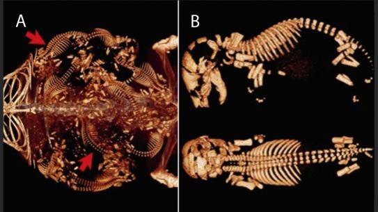
In addition, to his own research, Dr. Wadghiri leads the NYU Langone’s Preclinical Imaging Laboratory, which provides investigators with access to state-of-the-art imaging technologies and strategies to image live animals on the organ, tissue, cell, or molecular level. The available technologies are indispensable to researchers studying and monitoring disease in small living subjects. Built in partnership with the Department of Radiology, the newly renovated, 4,000-square-foot facility opened in May 2016 and enables researchers to perform noninvasive and nonlethal three-dimensional imaging. The lab is equipped with powerful instruments for micromagnetic resonance imaging (MRI), micropositron emission tomography, X-ray microcomputed tomography, bioluminescence and fluorescence scanning, and high-frequency ultrasound. The imaging instruments are located conveniently throughout NYU Langone’s facilities on First Avenue in Manhattan. Devices are located either within or outside the pathogenic barrier and near the various vivaria, providing expanded access and support to investigators conducting diverse biomedical research projects.


