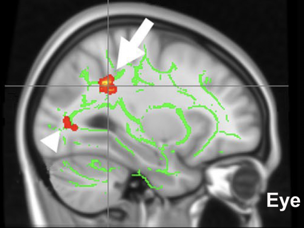
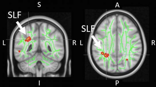
Structural, Metabolic and Functional Brain Imaging for Disease Detection and Therapeutic Monitoring in Glaucoma

Speaker:
Kevin C. Chan, PhD
Assistant Professor of Ophthalmology and Radiology
NYU Grossman School of Medicine
Abstract:
Glaucoma, the world’s leading cause of irreversible blindness, is a condition for which elevated intraocular pressure is currently the only modifiable risk factor. However, the disorder can continue to progress even at reduced intraocular pressure. This indicates additional key factors that contribute to the etiopathogenesis. There has been a growing amount of literature suggesting glaucoma as a neurodegenerative disease of the visual system. However, it remains debatable whether the observed pathophysiological conditions are causes or consequences. This review summarizes recent in vivo imaging studies that helped advance the understanding of early glaucoma involvements and disease progression in the brains of humans and experimental animal models. In particular, we focused on the non-invasive detection of early structural and functional brain changes before substantial clinical visual field loss in glaucoma patients; the eye-brain interactions across disease severity; the metabolic changes occurring in the brain’s visual system in glaucoma; and, the widespread brain involvements beyond the visual pathway as well as the potential behavioral relevance. If the mechanisms of glaucomatous brain changes are reliably identified, novel neurotherapeutics that target parameters beyond intraocular pressure lowering can be the promise of the near future, which would lead to reduced prevalence of this irreversible but preventable disease.


