
Proton Magnetic Resonance Spectroscopic Imaging in Clinical Applications and Neuroscience Research
Speaker:
Dikoma C. Shungu, Ph.D.
Professor of Physics in Radiology
Director, Laboratory for Advanced MRS Research
Weill Cornell Medical College
Abstract:
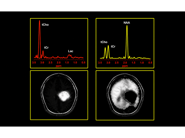
Magnetic resonance imaging (MRI), now an established noninvasive neuroimaging modality for clinical evaluation and monitoring of the therapeutic response of intracranial lesions, is not without limitations: the technique often lacks the specificity to differentiate pathologic lesions (e.g., brain tumors) from normal post-operative or post-therapy changes, such as edema and radiation-induced necrosis, and it can miss pathologic changes that do not exhibit contrastenhancement (e.g., low-grade brain neoplasms). As a result, there has been a great deal of interest in evaluating proton magnetic resonance spectroscopy (1H MRS) as a complement to contrast-enhanced MRI in the evaluation of intracranial lesions. 1H MRS is a noninvasive metabolic imaging technique that is closely related to MRI and can be performed in conjunction with MRI on virtually every clinical MRI scanner without the need to change hardware or move the patient. However, whereas MRI uses tissue water to produce highly detailed anatomic images, 1H MRS is concerned with the noninvasive assessment of neurochemistry by measuring the spatial distribution and concentrations of several endogenous compounds (e.g., amino acids, neurotransmitters) of biomedical interest. In this presentation, we will describe our experience using 1H MRS imaging (1H MRSI) as a powerful noninvasive complement to structural MRI for the diagnostic evaluation of intracranial lesions and as an aid to clinical decision-making. In addition, examples will be presented to demonstrate the utility of 1H MRSI as a general and powerful noninvasive tool for neuroscience research.