Tailored Single-Molecule Biophotonics Approaches for Targeted Development of Cancer Therapeutics and Treatment of Pain
Speaker:
Eli Rothenberg, PhD
Associate Professor, Biochemistry and Molecular Pharmacology
NYU Grossman School of Medicine
Abstract:
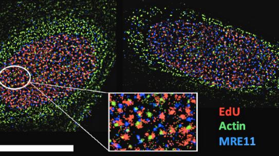
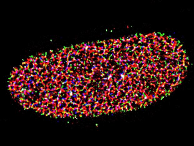
Single-molecule probing methods provide unique information that cannot be obtained with conventional ensemble techniques due to averaging. These cutting-edge techniques offer new probing capabilities to monitor dynamics of individual proteins and complexes in real time, and image molecules in cells with a resolution of several nanometers. These methods are conceptually groundbreaking and extremely promising in providing a plethora of new information and shedding light on major biological questions. Dr. Rothenberg’s group is developing and using state-of-the-art single-molecule fluorescence imaging techniques. This includes methods such as multi-color single-molecule localization microscopy, single-molecule FRET, and single-molecule tracking. They use these tools to study specific molecular features and mechanisms of biological systems.
For his BS degree Physical Chemistry, Dr. Rothenberg performed research at Farkas Center for Light Induced Processes and the Center for Nanoscience and Nanotechnology at the Hebrew University of Jerusalem in Israel. Subsequently, he pursued a degree in physics under the tutelage of Prof. Sevin at the University of Illinois at Urbana-Champaign. During his PhD he focuses on novel fluorescence resonance energy transfer (FRET) methods for single molecule imaging at the Center for Physics of the Living Cell. In 2011 he accepted an offer for an Assistant Professor position at the Dept. of Biochemistry and Molecular Pharmacology at NYU. Here he continued his development of cutting-edge single-molecule fluorescence imaging techniques and their use for studying molecular mechanisms relevant to diverse human diseases and syndromes.